


- 相關法規 -
18.Jul.2008
X-ray螢光-XRF 環境檢測方法─ EPA 6200 Method 標準土壤底泥檢測方法

環境檢測方法─ EPA 6200 Method標準土壤底泥檢測方法
根據美國環境保護署(美國環境保護局)的EPA 6200 Method,以攜帶式X - 射線螢光光譜儀(Field portable X - ray fluorescence,以下簡稱 FPXRF)來分析並推估土壤和汙泥中元素濃度。6200 Method法檢測土壤和底泥中 26 種金屬元素之偵測限值。有些常見的元素沒有被列在其中,因為它們是屬於”輕(Light)”的元素,無法使用 FPXRF 來分析,這些元素包括鋰、鈹、鈉、鎂、鋁、矽和磷。一般原子序在 16 以上的元素皆可使用 FPXRF 儀器來檢測和定量。
XRF原理
X-光螢光分析儀 (X-ray Fluorescence Spectrometer, XRF )係利用X-光束照射試片以激發試片中的元素,當原子自激發態回到基態時,偵測所釋放出來的螢光,經由分光儀分析其能量與強度後,可提供試片中組成元素的種類與含量,具有快速、非接觸、非破壞性及多元素分析等特點。
X-光螢光分析儀 (X-ray Fluorescence Spectrometer, XRF )係利用X-光束照射試片以激發試片中的元素,當原子自激發態回到基態時,偵測所釋放出來的螢光,經由分光儀分析其能量與強度後,可提供試片中組成元素的種類與含量,具有快速、非接觸、非破壞性及多元素分析等特點。
其原理是利用放射源(如Cd109)或高電壓激發光管(非輻射放射源)放射出X-ray,激發待測物內層的電子,此時為保持能量平衡,分子外圍電子補入內層電子的空缺。因外圍電子能量較高,故外為電子進入內層軌域時,放出特定的螢光能量,藉由能量的大小推知待測物的量。
一. 設備與材料
(一)FPXRF 光譜儀:
一般而言 FPXRF 光譜儀應至少包含四個主要組件,以下針對各組件作一概述:
1.X -射線放射源:產生 X - 射線的放射源,如以放射性同位素放射源(如 Fe - 55、Cd - 109、Am – 241 或Cm – 244 等)或以 X - 射線管(以高電壓加速電子撞擊陽極靶材如銅、鉬或銀等產生電子激發)當作 X - 射線的放射源。
2.樣品承接裝置:現場直接檢測或以樣品採集後再放置在樣品承裝器中分析。
3.X - 射線偵測器:包括固態式偵測器或氣體填充式比例計數偵測器等。固態式偵測器包括碘化汞(HgI2)、矽汞和鋰矽Si(Li),碘化汞(HgI2)偵測器可使用低必v熱電式冷卻裝置來控制操作溫度在低於室溫下,矽汞偵測器也可經由熱電式Peltier效應來冷卻之,而鋰矽偵測器則必需被冷卻至最少 - 90 ℃,其冷卻方式可使用液態氮或經由熱電式 Peltier 效應來冷卻之。
4.數據處理系統:包括測量脈衝振幅之多頻路分析儀(MCA),可將訊號處理成 X - 射線能量光譜,然後再計算出樣品中元素濃度的分析儀,及分析數據顯示和儲存系統。
(二)備用電池或充電器。
(三)聚乙烯(Polyethylene,PE)製樣品承裝器:直徑 31 mm 到 40 mm,有軸環或同級品(適用在 FPXRF 儀器)。
(四)X -射線端視窗上用之薄膜:MylarTM、KaptonTM、SpectroleneTM、Polypropylene 或同級品;厚度 2.5 至 6.0 μm。
(五)研缽和碾槌:玻璃,瑪瑙,或氧化鋁材質,用來磨碎土壤和底泥樣品。
(六)樣品瓶:玻璃或塑膠製品,用來貯存樣品。
(七)篩網:60 篩目(0.25 mm),不鏽鋼,尼龍或同級品,用來製備土壤和底泥樣品。
(八)小鏟子:用來弄平土壤表面及採集土壤樣品用。
(九)塑膠袋:承裝土壤及混合土壤使之均勻化。
(十)烘箱:烘乾土壤和底泥樣品用的烘箱或烤箱。
二. SUMMARY OF METHOD
1. The FPXRF technologies described in this method use either sealed radioisotope sources or x-ray tubes to irradiate samples with x-rays. When a sample is irradiated with x-rays, the source x-rays may undergo either scattering or absorption by sample atoms. This latter process is known as the photoelectric effect. When an atom absorbs the source x-rays, the incident radiation dislodges electrons from the innermost shells of the atom, creating vacancies. The electron vacancies are filled by electrons cascading in from outer electron shells. Electrons in outer shells have higher energy states than inner shell electrons, and the outer shell electrons give off energy as they cascade down into the inner shell vacancies. This rearrangement of electrons results in emission of x-rays characteristic of the given atom. The emission of x-rays, in this manner, is termed x-ray fluorescence.
Three electron shells are generally involved in emission of x-rays during FPXRF analysis of environmental samples. The three electron shells include the K, L, and M shells. A typical emission pattern, also called an emission spectrum, for a given metal has multiple intensity peaks generated from the emission of K, L, or M shell electrons. The most commonly measured x-ray emissions are from the K and L shells; only metals with an atomic number greater than 57 have measurable M shell emissions.
Each characteristic x-ray line is defined with the letter K, L, or M, which signifies which shell had the original vacancy and by a subscript alpha (α), beta (β), or gamma (γ) etc., which indicates the higher shell from which electrons fell to fill the vacancy and produce the x-ray. For example, a Kα line is produced by a vacancy in the K shell filled by an L shell electron, whereas a Kβ line is produced by a vacancy in the K shell filled by an M shell electron. The Kα transition is on average 6 to 7 times more probable than the Kβ transition; therefore, the Kα line is approximately 7 times more intense than the Kβ line for a given element, making the Kα line the choice for quantitation purposes.
The K lines for a given element are the most energetic lines and are the preferred lines for analysis. For a given atom, the x-rays emitted from L transitions are always less energetic than those emitted from K transitions. Unlike the K lines, the main L emission lines (Lα and Lβ) for an element are of nearly equal intensity. The choice of one or the other depends on what interfering element lines might be present. The L emission lines are useful for analyses involving elements of atomic number (Z) 58 (cerium) through 92 (uranium).
An x-ray source can excite characteristic x-rays from an element only if the source energy is greater than the absorption edge energy for the particular line group of the element, that is, the K absorption edge, L absorption edge, or M absorption edge energy. The absorption edge energy is somewhat greater than the corresponding line energy. Actually, the K absorption edge energy is approximately the sum of the K, L, and M line energies of the particular element, and the L absorption edge energy is approximately the sum of the L and M line energies. FPXRF is more sensitive to an element with an absorption edge energy close to but less than the excitation energy of the source. For example, when using a cadmium-109 source, which has an excitation energy of 22.1 kiloelectron volts (keV), FPXRF would exhibit better sensitivity for zirconium which has a K line energy of 15.77 keV than to chromium, which has a K line energy of 5.41 keV.
2. Under this method, inorganic analytes of interest are identified and quantitated using a field portable energy-dispersive x-ray fluorescence spectrometer. Radiation from one or more radioisotope sources or an electrically excited x-ray tube is used to generate characteristic x-ray emissions from elements in a sample. Up to three sources may be used to irradiate a sample. Each source emits a specific set of primary x-rays that excite a corresponding range of elements in a sample. When more than one source can excite the element of interest, the source is selected according to its excitation efficiency for the element of interest.
For measurement, the sample is positioned in front of the probe window. This can be done in two manners using FPXRF instruments, specifically, in situ or intrusive. If operated in the in situ mode, the probe window is placed in direct contact with the soil surface to be analyzed. When an FPXRF instrument is operated in the intrusive mode, a soil or sediment sample must be collected, prepared, and placed in a sample cup. The sample cup is then placed on top of the window inside a protective cover for analysis.
Sample analysis is then initiated by exposing the sample to primary radiation from the source. Fluorescent and backscattered x-rays from the sample enter through the detector window and are converted into electric pulses in the detector. The detector in FPXRF instruments is usually either a solid-state detector or a gas-filled proportional counter. Within the detector, energies of the characteristic x-rays are converted into a train of electric pulses, the amplitudes of which are linearly proportional to the energy of the x-rays. An electronic multichannel analyzer (MCA) measures the pulse amplitudes, which is the basis of qualitative x-ray analysis. The number of counts at a given energy per unit of time is representative of the element concentration in a sample and is the basis for quantitative analysis. Most FPXRF instruments are menu-driven from software built into the units or from personal computers (PC).
The measurement time of each source is user-selectable. Shorter source measurement times (30 seconds) are generally used for initial screening and hot spot delineation, and longer measurement times (up to 300 seconds) are typically used to meet higher precision and accuracy requirements.
FPXRF instruments can be calibrated using the following methods: internally using fundamental parameters determined by the manufacturer, empirically based on site-specific calibration standards (SSCS), or based on Compton peak ratios. The Compton peak is produced by backscattering of the source radiation. Some FPXRF instruments can be calibrated using multiple methods.
資料來源
美國環境保護署
美國環境保護署
XRF 在釵h研究報告中已被證實為有效的「篩選工具」,已漸漸的替代傳統化學分析方式,而成為主要「分析工具」之一。科邁斯集團對於XRF 製造商也積極對軟硬體技術、標準樣品進行研發新的設備,針對「取樣點的選擇」、「取樣數量」及「數據可信度」做更廣泛的研究與討論定義新的測試方法。

其他相關訊息
-
 22.Apr.2024
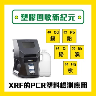
X-ray螢光-XRF 塑膠回收新紀元:XRF在PCR塑料檢測的創新應用
-
 04.Mar.2024
X-ray螢光-XRF 原能會改制? 別擔心,科邁斯一站式服務讓您無後顧之憂!
-
 26.Feb.2024
X-ray螢光-XRF 面對RoHS 3.0新添限制物質該如何對應?